Meet the International Medical Imaging Expert Improving Cancer Patient Outcomes
As part of UC Davis Global Affairs, Services for International Students and Scholars (SISS) is helping to build a campus community that includes students and scholars from over 100 countries and six continents. Each year, SISS serves more than 10,000 international students, faculty and researchers and their accompanying family members who come to UC Davis.
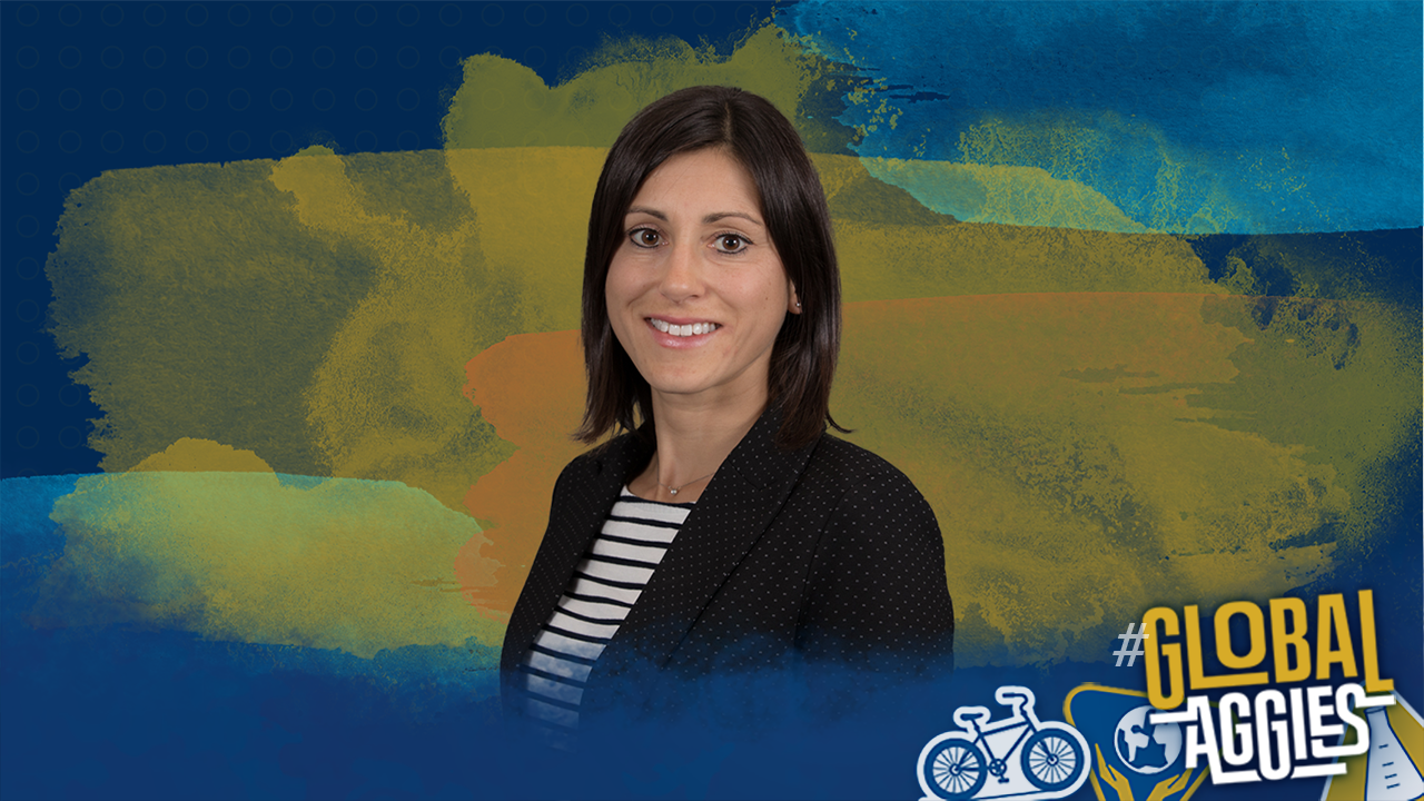
With expertise in medical imaging, Emilie Roncali is one such international scholar.
A leading biomedical engineer and physicist, Roncali focuses on medical imaging and the effectiveness of medical treatments for cancer patients. Her work involves molecular imaging and therapy, with an emphasis on new technology for positron emission tomography (PET).
“Simply put, I am working on nuclear medicine technology, improving diagnostic medical imaging devices and also studying radiation dose in radiation therapy for cancer patients,” she said.
An International, Interdisciplinary Journey
Born and raised in France, Roncali came to the United States for a postdoctoral research position in the Cherry Lab at UC Davis, run by Distinguished Professor Emeritus Simon Cherry.
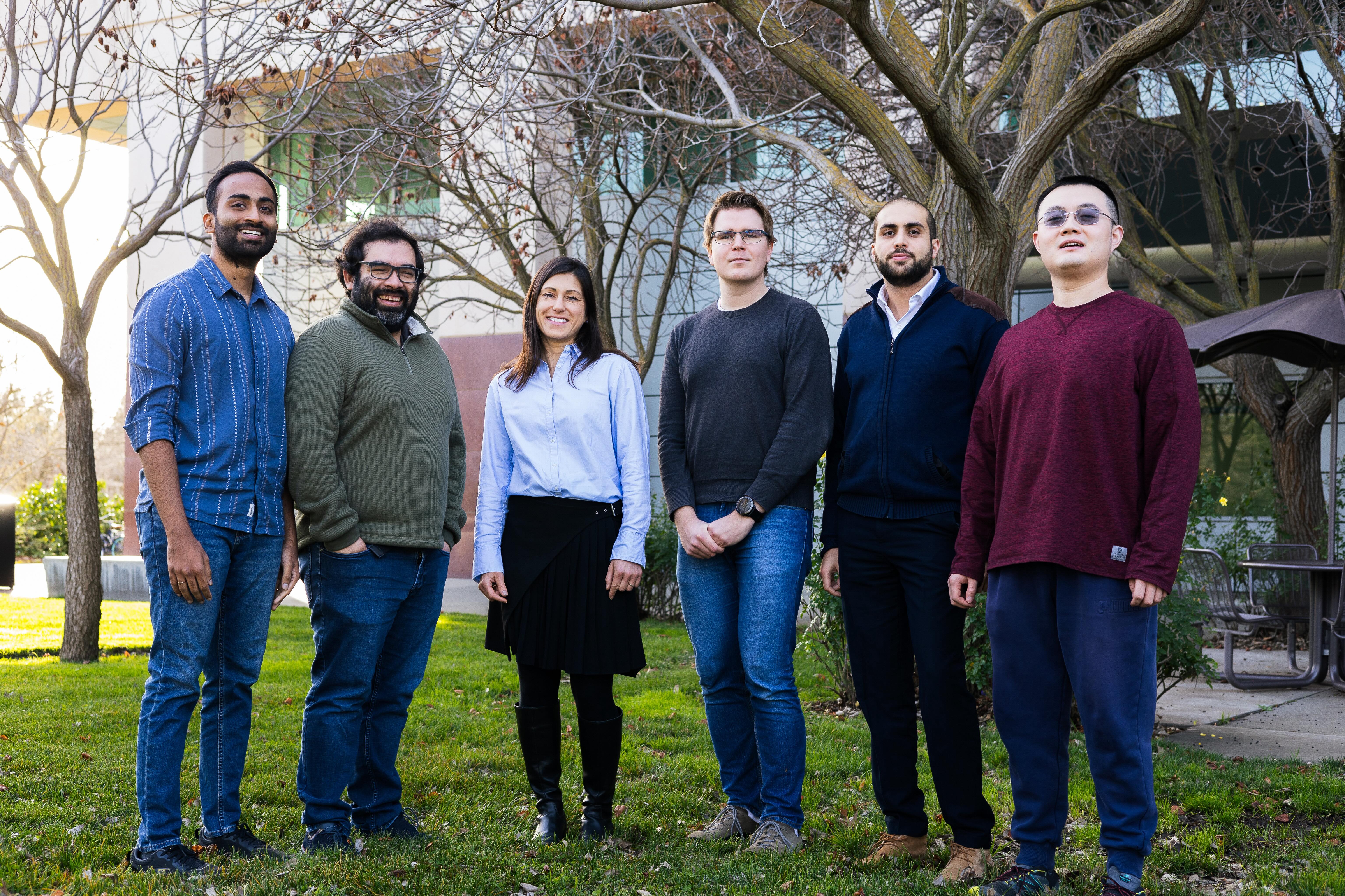
Now the head of her own group, in the Roncali Lab, she and her international team of postdoctoral researchers and students focus on modeling and simulation for medical imaging and therapy physics. They apply a biomedical engineering perspective to solve clinical problems.


“While our lab is housed in the Department of Biomedical Engineering, we recruit students and postdocs in mechanical engineering, computer science, electrical engineering—and, importantly, medical students from UC Davis Health,” she said. “This makes our work incredibly interdisciplinary, and we take on many multidisciplinary projects.”
Roncali said her lab is currently developing new methods to calculate the radiation dose before and after cancer treatment. This means that her team can assess the treatment's efficacy and the toxicity. Ultimately, the work helps physicians better personalize treatment to achieve longer overall survival for cancer patients, while also improving their quality of life.
“By leading a team of international researchers, I can observe and harness the power of different thinking processes and learn from their work regime on a daily basis, essentially helping them while they help us.”
Roncali has a joint faculty appointment with the Department of Biomedical Engineering at UC Davis and the Department of Radiology at UC Davis Health. In addition to her responsibilities at UC Davis, she is a member of two focus groups for the National Cancer Institute as well as an investigator in the Informatics Technology for Cancer Research initiative, to improve treatment planning and optimize testosterone replacement therapy in clinical trials.
In 2022, she was awarded the Tracy Lynn Faber Memorial Award from the Society of Nuclear Medicine and Molecular Imaging for her outstanding contributions to advancing women in the medical imaging sciences. The award is given to an individual who has significantly promoted the advancement of women in medical imaging sciences or to a woman in early- or mid-career who has made one or more significant contributions to medical imaging sciences.
Supporting Students and the Global Research Community
Roncali’s medical imaging simulation tools are frequently released for free in open-source software. These tools are broadly used in nuclear medicine technology development and help many scientists who would not otherwise have access to such sophisticated systems.
“Our current partner for a project on AI for medical imaging simulations is the National Centre for Scientific Research, based in Lyon, France,” she said. “Two of my trainees are very active with that group in France, and we collaborate with many international colleagues.”
In addition to running a lab, mentoring students and teaching imaging physics to physicians, Roncali also teaches undergraduate courses on probability and data science for biomedical engineers and graduate courses on nuclear imaging in medicine and biology.
“For my undergraduate students, I hope to convince them that probability is a useful tool in biomedical engineering and is everywhere in life and physics,” she said. “For my graduate students, I hope they understand the direct application of physics to clinical imaging and connect the science to the application in their future work as scientists and researchers.”
Thanks to a track record of establishing new research projects and securing internal and external funding, Roncali has made it her professional mission to address unmet clinical imaging and therapy needs.
“My ultimate aim is to improve cancer patient outcomes and advance cancer research through personalized medicine,” she said.